
The Physiology of Hearing
Introduction
The physiology of our hearing mechanism can conveniently be divided into three topics:
1 The outer ear (auricle or pinna) and ear canal
2 The middle ear
3 The inner ear
The Auricle and Ear Canal.
Each hole in the side of the skull leads into an ear canal. The ear canal is an irregular cylinder with an average diameter of less than 0.8 mm and about 2.5 cm long.

The ear canal (figure 1) is open at the outer end which is surrounded by the pinna (or auricle). The pinna plays an important spacial focusing role in hearing. The canal then narrows slightly and widens towards its inner end, which is sealed off by the eardrum.
Thus the canal is a shaped tube enclosing a resonating column of air - with the combination of open and closed ends. This makes it rather like an organ pipe.
The ear canal supports (resonates or enhances) sound vibrations best at the frequencies which the human ears hear most sharply. This resonance amplifies the variations of air pressure that make up sound waves, placing a peak pressure directly at the eardrum.
For frequencies between approximately 2 KHz and 5.5 KHz, the sound pressure level at the eardrum is approximately 10 times the pressure of the sound at the auricle.
The Eardrum - interface between outer and middle ear.
Airborne sound waves reach only as far as the eardrum. Here they are converted into mechanical vibrations in the solid materials of the middle ear.
Sounds (air pressure waves) first set up sympathetic vibrations in the taunt membrane of the eardrum, just as they do in the diaphragm of some types of microphone. The eardrum passes these vibrations on to the middle ear structure.
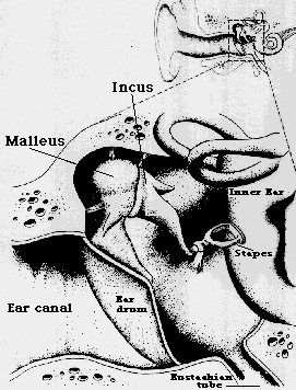
The Middle Ear Ossicular Chain - Malleus (Hammer) , Incus (Anvil) , and Stapes (Stirrup).
The middle ear contains three small bones known as the Malleus, Incus, and Stapes. (Fig. 2). These bones form a system of levers which are linked together and driven by the eardrum. Malleus pushing Incus, Incus pushing Stapes.
Working together as a lever system, the bones amplify the force of sound vibrations.
The inner end of the lever moves through a shorter distance but exerts a greater force than the outer end.
In combination the bones double or triple the force of the vibrations at the eardrum.
The muscles of the middle ear modify the performance of this lever system as an amplifying unit. They act as safety devices to protect the ear against excessively large vibrations from very loud sounds - a sort of automatic volume control.

Although these tiny muscles - the smallest in the human body - at both ends of the ossicular chain can be moved voluntarily, their normal contractions are reflex triggered when sound exceeds a certain level. As the noise level rises, one set of muscles tightens to restrict the movement of the malleus thus weakening the vibrations transmitted within the middle ear. At the same time the stapes muscle contracts to pull the stapes away from the oval window so that less vibration is passed along to the very sensitive inner ear.
The inner end of the lever moves through a shorter distance but exerts a greater force than the outer end.
In combination the bones double or triple the force of the vibrations at the eardrum.
The muscles of the middle ear modify the performance of this lever system as an amplifying unit. They act as safety devices to protect the ear against excessively large vibrations from very loud sounds - a sort of automatic volume control.
Although these tiny muscles - the smallest in the human body - at both ends of the ossicular chain can be moved voluntarily, their normal contractions are reflex triggered when sound exceeds a certain level. As the noise level rises, one set of muscles tightens to restrict the movement of the malleus thus weakening the vibrations transmitted within the middle ear. At the same time the stapes muscle contracts to pull the stapes away from the oval window so that less vibration is passed along to the very sensitive inner ear.
The Oval Window - Interface Between Middle & Inner Ear.
The stirrup passes the vibrations to the "oval window" which is a membrane covering an opening in the bony case of the cochlea.
The size of the oval window compared to that of the eardrum (15 to 30 times smaller) produces the critical amplification needed to match impedances between sound waves in the air and in the cochlear fluid. Apart from the amplification of the bone lever system of the middle ear, this concentration of force amplifies the incoming vibrations of sound about 15 to 30 times.
Summary of the Amplifications from Outer to Inner Ear.
Within the 4 cm or so occupied by the outer and middle ears, three distinct physical principles operate to magnify weak vibrations in air so that they can establish pressure waves in a liquid:
- The organ pipe resonance of the ear canal may increase the air pressure fore 10 times.
- The mechanical advantages of the bone lever system may nearly triple it.
- The pinpointing arrangement of the eardrum and the oval window may provide another thirty fold increase.
The result of these three mechanisms may be an amplification of a sound wave by more than 800 times before it sets the liquid of the inner ear in motion.
The Inner Ear : The Cochlea.
The amplified mechanical force transmitted from the middle ear to the inner ear by the ossicles is immediately transformed in the cochlea into hydraulic pressure. This hydrolic pressure imparts movement to the cochlear duct and to the organ of Corti - the "peripheral seat of hearing". This process is acomplished in the cochlea which is no bigger than the tip of a little finger.
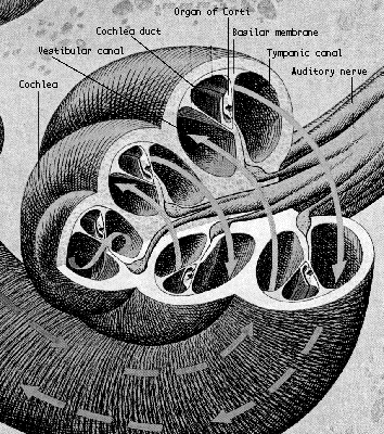
A cross-section of the cochlea shows its vestibular canal, tympanic canal and cochlear duct.
The vestibular and tympanic canals contain perilymph - a liquid almost identical with spinal fluid. The cochlea duct contains endolymph - a liquid similar to the fluid within cells. The canals are separated by thin membranes: Reissner's membrane between the vestibular canal and the cochlea duct is the thinnest (just 2 cell walls thick), and the basilar membrane between the tympanic canal and the cochlear duct.
The perilymph and endolymph have differing chemical compositions and electrical charges. Any break in the membranes that allows them to mix impairs the hearing process.
How the Cochlea works.
The pressure waves in the cochlea exert energy along a route that begins at the oval window and ends abruptly at the membrane-covered round window, where the pressure is dissipated. Inkeeping with the principles of hydraulics, the pressure applied to the oval window at the stirrup is transmitted to all parts of the cochlea.
The semicircular canals are the body's balance mechanism and it is thought that it plays no part in hearing.
One of the two walls of the cochlear duct is the vibrating basilar membrane whose function is to separate sounds according to frequency. The membrane is narrow and taunt at the end near the stirrup and wider and more pliant at the other.
Hydraulic pressure waves in the cochlea induce a wave-like ripple in the basilar membrane which travels from the taunt towards the loose end. High tones create their greatest crests where the membrane is tight, low tones where the wall is slack. This is because resonant frequency is correlated with tension as in a taunt string. The position of this crest is important because it determines which nerve fibres will send signals to the brain. High frequency tones cause the crest to occur at the base of the cochlea and the lower frequencies towards the apex.
Apart from airborne sounds, the basilar membrane also picks up vibrations in the skull from such sources as teeth clicking, for example.
The Organ of Corti.
The Organ of Corti is a gelatinous mass about 4 cm long and is composed of some 7500 interrelated parts.The Organ of Corti is enclosed in the cochlea which is deeply imbedded in the temporal bone (the hardest in the body) is one of the best protected parts of the body. It is related to a series of tiny sensing bumps in fishes that are located along the body in rows just under the skin. These tiny bumps are used by fish to sense slight movements of water. The Organ of Corti operates in a similar way. It is filled with fluid, surrounded by other fluid and responds to movements in these fluids - those movements induced by sound waves.
The fluids filling and surrounding it act as shock absorbers, and so do the springy membranes which support it. It is even isolated from the normal body supply lines, for the faint pulsing of blood through capillary vessels would be detected as background noise. The capillaries nearest to the organ of Corti end at the wall of the cochlea; nutrients on their way out are carried to and from the capillaries by the endolymph fluid that bathes the organ.
The organ of Corti is shaped like the jam in a jam roll. It spirals around within the cochlea. The basilar membrane supports the organ which contains a mass of cells almost touching the branch endings of the auditory nerve. From these cells sprout fine hairs, (23,500 of them) rising in orderly rows like the bristles of a very soft brush. The hairs stick through the dome of the organ, their ends embedded in a thick overhanging sheet, the tectorial membrane. These hairs are transducers. As the basilar membrane bellies in and out, it pushes and pulls the complex of tissues above it. The hairs' cells of the organ of Corti ride with the basilar membrane. The hairs have their tops embedded in the tectorial membrane and their roots fixed in the hair cells, so the motion of the basilar membrane bends and twists and pulls and pushes the hairs. Under these physical stresses the hairs generate electrical signals which stimulate the auditory nerve (also known as the acoustic nerve and the eighth cranial nerve) - a bundle of about 30,000 individual fibres.
Eventually, in a way still not fully understood, the electrical signals running through the auditory nerve stimulate the hearing centres of the brain. In the cells of the auditory cortex lies the mystery of the sensation of hearing.
The organ of Corti serves two vital functions:
- It Converts mechanical energy into electrical energy.
- It Dispatches to the brain a coded version of the original sound - information not only about fundamental frequency but about intensity and timbre as well.
As the organ of Corti, which is attached to the basilar membrane, bends to outside pressure, it moves laterally to the left whilst the tectorial membrane moves to the right.
This shearing action within the cochlear duct activates the hair cells of the organ of Corti to send their electrochemical signals into the central nervous system.
The Cochlea to brain transmission system.
The Cochlea to brain transmission system contains 30000 nerve fibres coming from the organ of Corti to form the auditory nerve. The fibres are grouped by the frequency of the sound signal they carry, the number of fibres a sound requires gives the brain a gauge of its intensity.
Coming from the brain, descending nerve fibres may carry instructions from the brain back to the ear to filter out and thus eliminate some signals which the brain determines are of no importance, and concentrate on others. After passing through the cochlear nucleus, some of the descending nerves from the brain go to the middle ear where they control the muscles used for fending off dangerously loud sound.
The nerve fibres carrying sound signals lead to different parts of the auditory cortex depending on the frequencies they carry. The auditory cortex lies in a deep furrow called the Sylvian fossa. The high tones terminate deep within the Sylvian fossa while the low tones end near the outer surface.
Postscript.
In traditional Chinese medicine, "The Kidneys open into the ear" ... There is a close relationship between the Kidneys and the ears. As Nei Jing says "The Kidney Qi goes through the ear; if the Kidney is harmonised, the ear can hear the five tones" Many hearing problems are treated through the Kidneys. The poor hearing common in the elderly, for example, is a consequence of weakened Kidney Jing.
Rigden, John S. :Physics and the Sound of Music: J. Wiley 2nd ed.: 1985
Blake, Randolph and Sekuler, Robert : Perception: Alfred A. Knopf, Inc. : 1985
Goldstein, E.Bruce : Sensation and Perception: Wadsworth Pub. Co. 3rd ed.: 1989
Stevens, S.S. & Warshofsky, Fred.: Sound and Hearing : Time-Life Science Library : 1980
Back to main course The Physics And Psychophysics Of Sound & Music
Back to Tuning, Scales And Temperament
Next to The Psychophysics of Hearing